Buyers and adopters of horses should learn as much as possible about the physical condition of the horses they are considering.
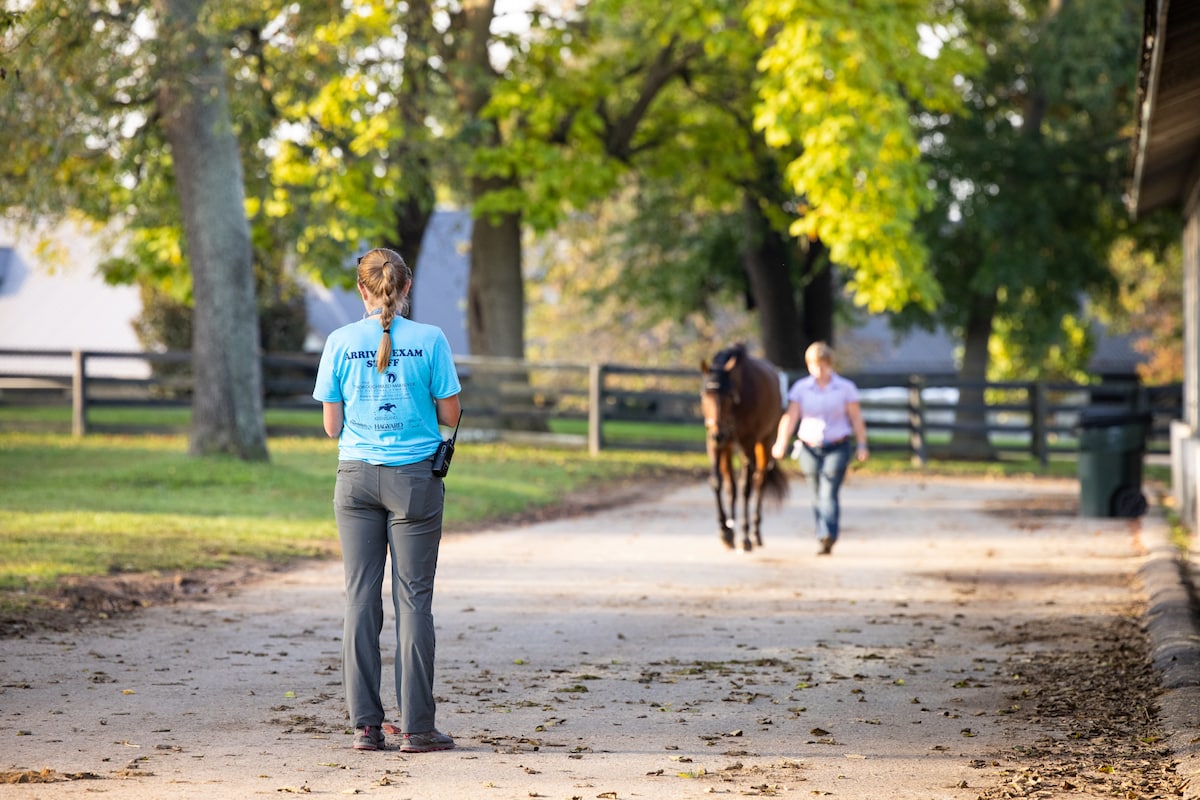
Bethany P Photography
The information below was compiled by Dr. Near specifically for the TCA Thoroughbred Marketplace, borrowing from a list of common injuries in racehorses on the Thoroughbred Owners and Breeders Association (TOBA) web site. Keep in mind that most horses, regardless of breed or discipline, have had injuries in the past and will continue to get them throughout their lives. Make your own decisions in consultation with experienced professionals about suitability and potential soundness, but keep in mind that most riding disciplines put much less stress on a horse’s limbs, heart, and lungs than racing. The soundness of the horses you are evaluating here has been tested.
Bog Spavin
Filling in the upper aspect of the hock joint. May be due to a variety of ailments (see OCD, bone chip).
Bone chip
As mentioned above, it is possible that joint filling (effusion) is due to the presence of a bone chip in the joint. These bone chips are small fragments off of the larger bones making up the joint and can either be free floating in the joint or loosely attached to other structures. The fragments can be traumatic or developmental in nature (see OCD). The free-floating fragments tend to be more problematic as they can erode or damage the articular cartilage resulting in osteoarthritis and lameness. The best way to determine the significance is with a soundness examination combined with radiographs of the joint. Prognosis for athletic function depends on the size and location of the chip as well as the joint affected.
Bone Spavin
Bone proliferation in the lower hock joints, which is seen as an increased profile on the inside of the lower aspect of the hock. Usually represents the chronic condition of the lower hock joints fusing and may cause lameness in the early stages.
Bowed Tendon
An inflammation and enlargement of the superficial or deep digital flexor tendon (located behind the cannon bone). The general cause is severe strain and the injury usually occurs in the front limbs. Back at the knees, long/weak pasterns, a long toe-low heel conformation and improper shoeing are all predisposing causes. The bowed appearance is due to inflammation followed by the formation of scar tissue. Bows are classified as low, medium or high depending on location. Treatment usually requires rest (6 months to a year). Afterwards, the tendon may continue to have an abnormal profile (old bow). Prognosis depends on the extent of the tendon damage but for most cases is quite good if rested sufficiently.
Bucked Shins
Inflammation of the tissue covering the front of the cannon bone, which can also extend into the cortex of the bone itself. Most frequently occurs in the front legs of young horses adjusting to the increased workload/concussion of training. Sometimes treated with pin-firing (resulting in a pattern of small round scars on the legs) and decreased workload. Prognosis for long-term soundness is good.
Condylar Fracture
A fracture of the condyle (the bulbous bottom or distal end of the cannon bone that fits into the fetlock joint). These more commonly occur in the front legs but can occur in the hind legs as well. Condylar fractures can be repaired surgically with screws placed across the fracture line or managed medically with bandaging and stall rest. In general, surgical repair leads to increased stability and thus decreased osteoarthritis of the fetlock joint, resulting in a better prognosis for long-term athletic function.
Curb
A firm enlargement on the rear of the cannon bone immediately below the hock. It begins as an inflammation of the plantar ligament and the inflammation leads to a thickening of the ligament. A sickle-hocked conformation can predispose this condition. Most often seen as a blemish and the prognosis for athletic soundness is very good.
Grabbed Quarter
While running, the horse “grabbed” one of its front hooves with a rear hoof, tearing skin and tissue. Lameness depends on the extent of the injury. Prognosis is very good.
Hygroma
A soft swelling most commonly found over the front of the knee that is outside of the joint capsule. The swelling represents accumulation of fluid in an acquired bursa, usually as the result of trauma. Usually seen as a blemish and the prognosis for athletic function is excellent.
Joint Effusion
Excessive synovial fluid or proliferation of the joint capsule can result in a joint being larger than usual. In OTTBs the joints most commonly affected are the fetlocks and knees. The large size of the joint is often indicative of underlying osteoarthritis, but can also result from the presence of loose bone fragments in the joint, inflammation, or chronic hyperextension (see Bone Chips, OCD, Osselets). The best way to determine the significance of the joint effusion is with a soundness examination combined with radiographs of the affected joint as needed.
OCD
Osteochondritis dissecans or OCD is a common developmental orthopedic disease of young horses, mainly affecting the articular cartilage and underlying bone in the fetlocks, hocks, and/or stifles. The main physical exam finding is increased fluid in the joint, although lameness may be present as well. Radiographs are necessary to determine the extent of the problem and may reveal a cystic lesion in the bone or loose fragments present in the joint. Treatment involves surgical removal of the loose fragments or rest and the use of chondroprotectants (i.e. Adequan) in the case of cystic lesions. Prognosis is variable depending on location and size of the lesion.
Osselets
A common term for osteoarthritis of the fetlock joint, most often represented as increased swelling located in the front of the joint. “Green osselets” (more acute cases) tend to be soft, where as chronic ones are more firm due to increased mineral deposition. Prognosis depends on the degree of osteoarthritis present (best evaluated using a soundness examination and radiographs).
Quarter Crack
A crack in the hoof wall located in front of the heels, which frequently results in unsoundness. Treatment involves stabilizing the crack and corrective shoeing. Prognosis is good aggressive treatment.
Sesamoid Fracture
The sesamoids are two small bones located at the back of the fetlock, held in place by a series of ligaments. The sesamoid bones are the main attachment of the suspensory ligament and also serve as pulleys over which the flexor tendons pass. Prognosis for athletic function depends on the location of the fracture and the extent of the suspensory damage.
Slab Fracture
A break in one of the main weight-bearing bones located in the horse’s knee whereby the “slab” of a carpal bone splits vertically and the front part becomes detached. This can often be repaired surgically (either by placing a screw across the fracture line or removing the detached part of bone if it is small) with a good prognosis or managed medically (stall rest) with a more guarded prognosis for athletic function (prognosis depends on the location of the fracture and the degree of displacement).
Splint
An increased calcification or bony growth of the splint bones (located on the inside and outside of the cannon bone). Most frequently occurring in the front limbs as a result of concussion with a hard surface or direct trauma. This injury typically results from a tear of the interosseous ligament that binds the splint bone to the cannon bone, but can result from traumatic fracture of the splint bone as well. Splints can be seen as an unsoundness (horse is often lame in the acute stages) or as a blemish (old splints may be unsightly but do not result in lameness). Prognosis for athletic function is excellent.
Suspensory Ligament
The suspensory ligaments run down the back of the cannon bone (deep to the flexor tendons) and attach on the sesamoid bones and pastern. These ligaments function to help suspend the fetlock joint. Injuries here are common in equine athletes. The treatment is usually six to nine months of rest with hand-walking. The prognosis for athletic function is fair to good depending on the location (front vs. hind leg, body vs. branch) and size of the suspensory injury.
Thoroughpin
Filling (excessive synovial or joint fluid) inside the synovial sheath of the flexor tendon located just above the hock. Most commonly seen as a blemish (horse is sound).
Windpuffs
Excessive fluid present in the tendon sheath at the level of the fetlock joint, most commonly in the hind legs. Usually represents a cosmetic blemish and does not result in lameness.
SEARCH
Sign up for our education newsletter
trending